For the first time, biologists managed to gain access to the sub-cellular world, via the use of an advanced algorithm and add-on technique, designed specifically for commercial microscope by researchers at the University of Illinois.
A paper detailing the study, entitled “Visualizing Escherichia coli sub-cellular structure using sparse deconvolution spatial light interference tomography,” is available in the June 29 online issue of the open-access journal PLoS ONE, which is edited by the Public Library of Science.
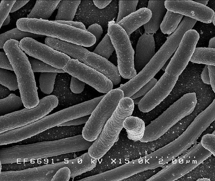
The microscopy method the team created can stealthily investigate the inner workings of the E. coli bacteria, and produces three-dimensional, high-quality images. Scientists believe that the approach may be used to analyze other types of living cells as well.
According to the PLoS ONE paper, the proof-of-concept study was carried out at great speed, scale and resolution, in a manner that cannot be replicated by any other label-free microscopy method currently available on the market.
While using contrast agents and fluorescent dyes continues to produce more in-depth results, the new approach may come in handy when scientists want to study a biological system without interference.
Gabriel Popescu, a researcher at the UIUC Beckman Institute for Advanced Science and Technology, says that the new microscopy method is based on a broadband interferometric technique known as Spatial Light Interference Microscopy (SLIM), which the expert himself designed.
SLIM works as an add-on module to a commercial phase contrast microscope, he reveals, and is capable of taking fast, accurate readings at several scales simultaneously, e! Science News reports.
“I believe this is a project that illustrates best the cross-pollination between different areas of expertise, which is so well nurtured at the Beckman Institute. We used a novel optical method in combination with advanced computer algorithms to tackle a problem of significance in biology,” Popescu says.
“This new method […] provides a way to take advantage of the intrinsic properties of these very small, transparent cells non-invasively and without the use of fluorescence techniques and contrast agents,” concludes UIUC researcher and first paper author, Mustafa Mir, a member of Popescu's research group.
Via: 3D Inner Structure of E. coli Resolved via Advanced Microscopy
Tidak ada komentar:
Posting Komentar